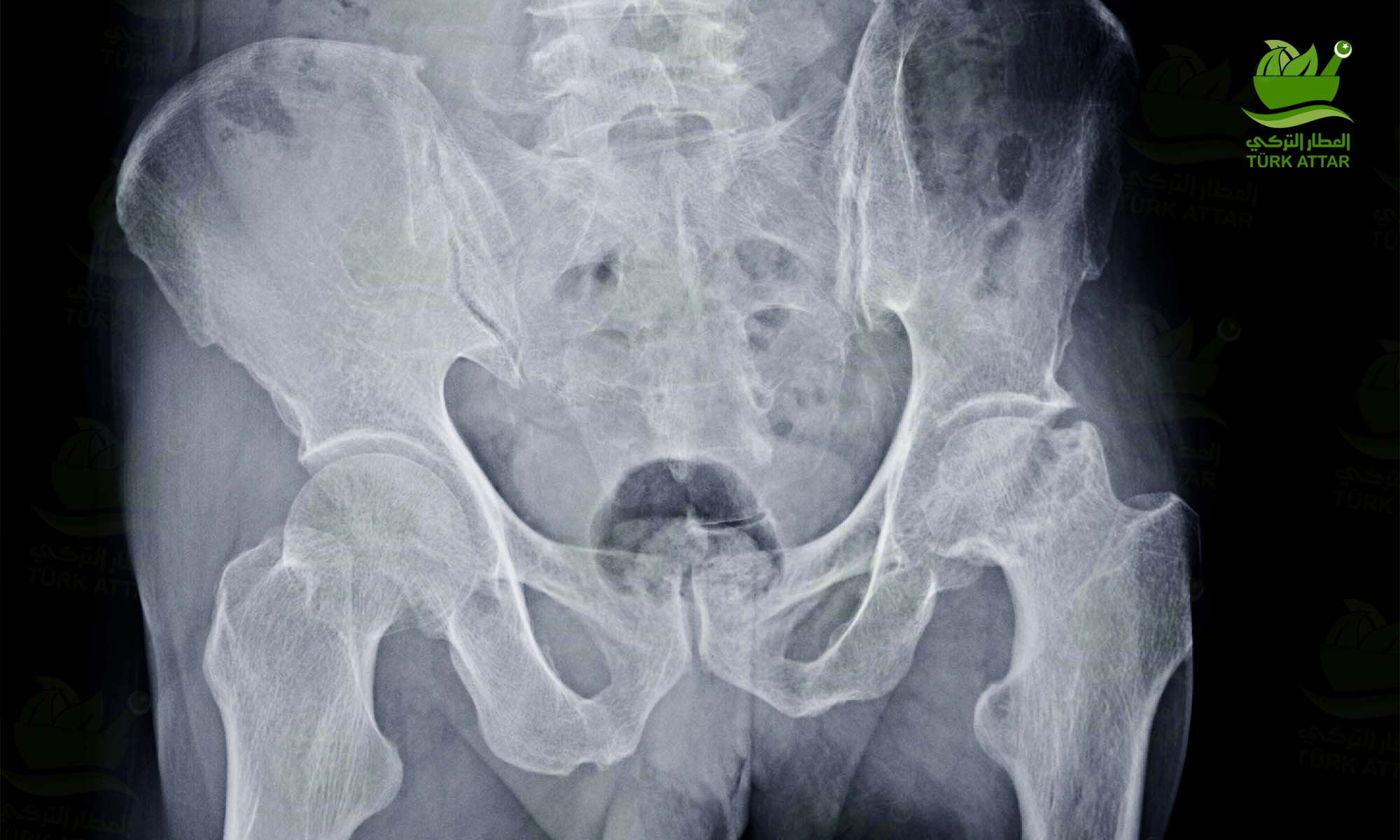
Avascular necrosis is most common in the hip joint. The blood circulation in the vessels feeding the femoral head stops and the bone under the articular cartilage begins to lose its vitality and collapses over time.
Avascular Necrosis
In fact, the most commonly known causes are; some blood diseases such as high-dose cortisone use, alcoholism, chemotherapy, cup coagulation disorders, sickle cell anemia.
Avascular Necrosis Symptoms and Diagnosis
The first symptom is groin pain. Although this pain can sometimes be so severe that it does not allow to step on it or even move the hip, there are also those who experience the process with mild pain. The two most typical features of the pain are that it increases with hip movements and spreads from the front of the leg to the knee. During the disease, painful and painless periods follow each other.
Diagnosis is made by examination and MRI. The biggest mistake made here is to send a person with hip pain to his/her home by taking an x-ray and saying it is not serious. Because up to the 4th stage, the x-ray can be seen as normal and valuable time is lost. As a rule, MRI is absolutely necessary for patients with hip pain and hip findings on examination.
MRI is 99% diagnostic. However, in some cases, it can be confused with a disease called temporary bone marrow edema. Inexperienced radiologists and orthopedists may cause unnecessary stress and surgery due to misdiagnosis.
In MRI, besides diagnosis, staging to determine treatment is also done. This requires a great deal of experience and mistakes to be made will result in wrong treatment.
Staging and Treatment
Stage I
There is edema under the articular cartilage in pain and MRI, but the edema boundaries are not clear. If the cause of the patient’s AVN can be known, it is concluded immediately. During this period, Core Decompression should be applied without losing any time. When done in this period, good results of up to 80% have been reported.
At this stage, ozone therapy, crutches, rest, hyperbaric oxygen applications are useless except for the loss of precious time.
Stage II
In MRI, the AVN area with clear borders is seen, but the cartilage is intact, there are no cracks and collapses. In this case, core decompression surgery should be performed without losing any time. Its success varies between 40-60%. Vascularized fibula graft surgery can be applied in patients under the age of 30 and in cases where core decompression does not work. It is successful around 80% in good and uncomplicated cases.
Stage III
At this stage, cracks and small collapses in the cartilage are seen on MRI. In this period, although there are those who make vascularized fibula grafts, there are also those who only treat pain. If the pain is very severe, a prosthesis can be made. I personally refer to the experienced microsurgery specialists for vascularized fibula grafts under the age of 30, stage III.
Stage IV
The load-bearing part of the head has collapsed. There is no treatment other than prosthesis. Interestingly, patients with less pain are also not few. Patients with less pain should wait for the prosthesis until the pain gets worse. Patients are not given any activity restrictions.
Core Decompression Surgery
The cessation of blood flow due to occlusion of the vessel in the femoral head first creates an edema. This edema increases the pressure in the femoral head, suppresses the blood flow in the non-occluded vessels, slows them down and even causes them to close with coagulation. Core decompression surgery technically consists of opening 3-4 3.2 mm or a single 8 mm tunnel into the edematous area. These tunnels or tunnels reduce the pressure caused by edema in the femoral head and allow the vessels under pressure to carry more blood. At the same time, these tunnels are perceived as broken and the inside of the tunnel is filled with new bone. New bone formation ensures the revival of the unnourished area due to both the rich vascular structure and abundant stem cells.
Only I and II. done in the universe. 80% in Stage I, II. 40-60% successful in the stage. Surgical quality is very important in success. In a quality surgery, tunnels or tunnels should be opened in the very center of the area that is completely closed under X-ray. This happens with both good operating room equipment and the cooperation of a patient and experienced surgeon.
Patients are given 25% weight bearing with double crutches for 6 weeks after surgery. They should use a single wand for 6-8 weeks.
Vascular Fibula Graft
The fibula is the thinnest of the two bones below the knee. There is no load bearing duty. 1/3 of this bone is taken from the middle of the bone by the artery and vein that feeds the bone. The fibula is placed in a hole drilled in the avascular femoral head. The veins are also stitched to the leg veins. Only experienced microsurgeons should perform this procedure. No matter how perfect the operation itself is, there is a risk of occlusion of the graft vessels after the procedure.

2 Comment(s)
1
1
1
1
1
Leave a Comment